Target
Where does the drug act?

Course orientation · Seven-step drug framework
A quick refresher—not a new framework
Where does the drug act?
What does it do there?
What changes directly?
How does the body respond?
Who benefits, and how?
What could go wrong, and why?
Does exposure change the prediction or decision?
We will highlight the one or two steps that do the most explanatory work for each drug, then reconnect them to the complete chain.
Foundations · 0.1
Event 1 of 11
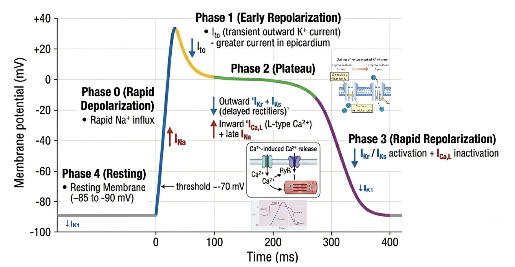
To Know Antiarrhythmics, You Have to Know the Action Potential
Atrial · ventricular · His–Purkinje
| Phase | Current | Event |
|---|---|---|
| 0 | INa | Upstroke. Rate of rise sets conduction velocity. |
| 1 | Ito | Notch. |
| 2 | ICaL vs IK | Plateau. Ca²⁺ entry triggers contraction. |
| 3 | IKr, IKs | Repolarization. Major determinant of APD and QT. |
| 4 | IK1 | Stabilizes rest; also contributes to terminal repolarization. |
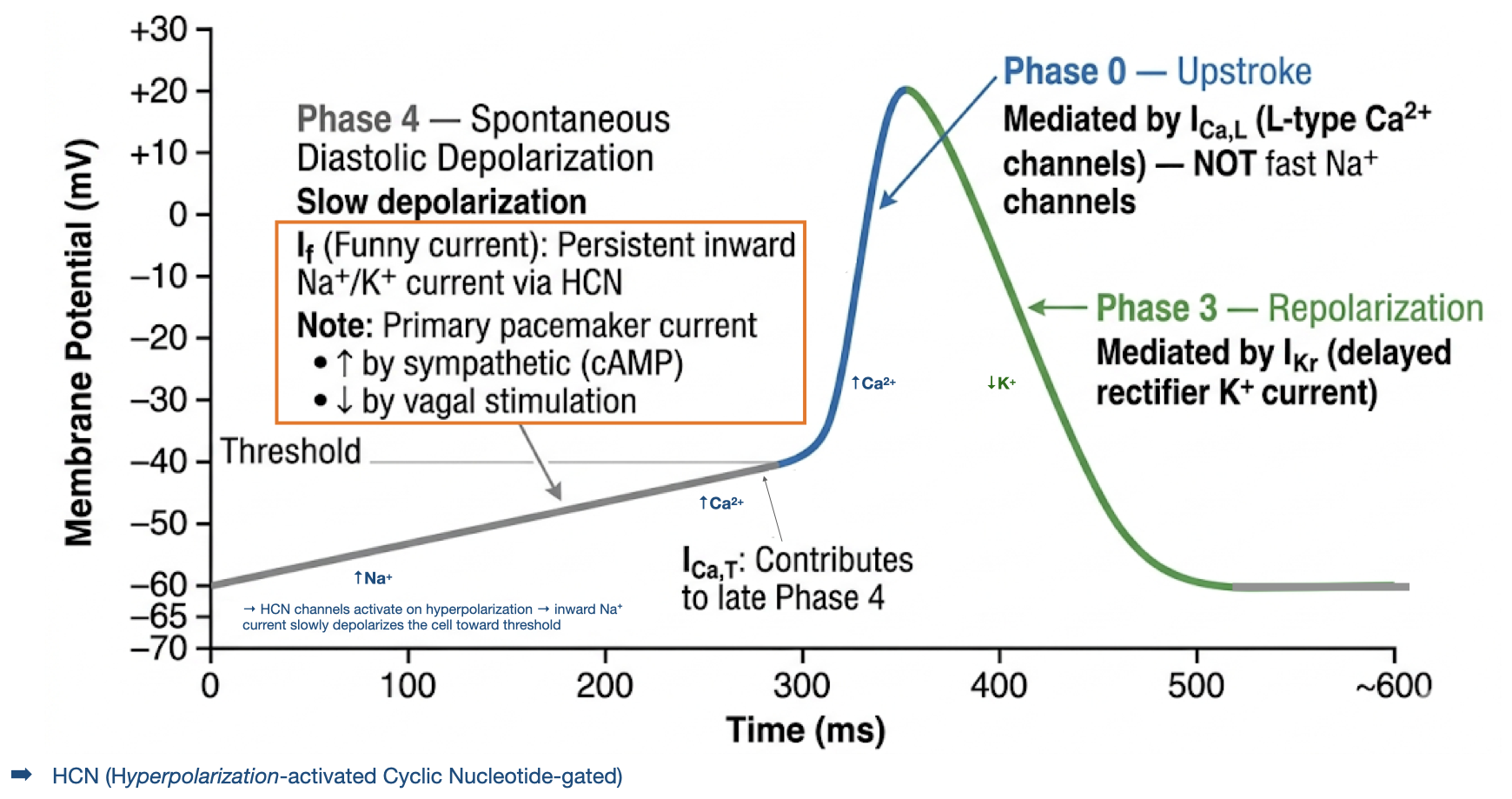
SA node · AV node
| Phase | Current | Event |
|---|---|---|
| 0 | ICaL | Slow upstroke; sets nodal conduction velocity. |
| 1 | — | Absent: no notch. |
| 2 | — | Absent: no plateau. |
| 3 | IK | Repolarization to maximum diastolic potential. |
| 4 | If, ICaT | Spontaneous depolarization toward threshold. |
Foundations · 0.2 · Channel states
Foundations · Mechanisms of arrhythmia
First ask what created the impulse. Then ask what allows it to continue.
Why did a cell fire?
Why did the wavefront conduct—or fail?
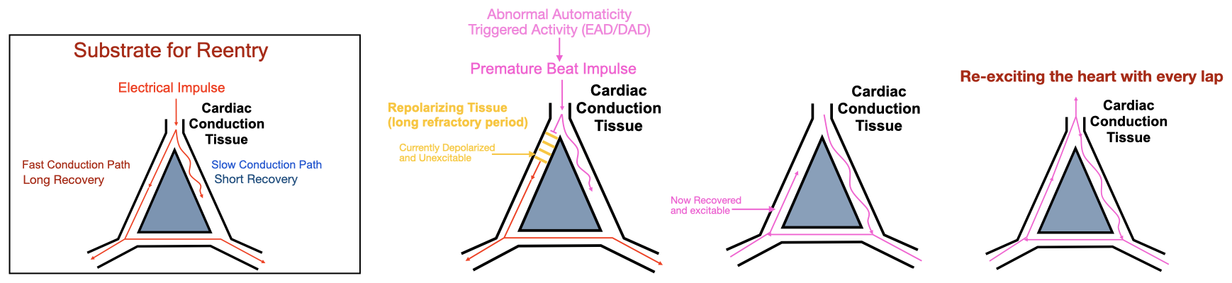
Foundations · 0.6 · Reentry
Anatomically or functionally distinct
One pathway blocks in one direction
The returning pathway recovers in time
Final synthesis · Step 1 study priorities
Separate fast-response atrial, ventricular, and Purkinje tissue from slow-response SA and AV nodal tissue.
Fast cells: Na⁺ phase 0, Ca²⁺ phase 2, and K⁺ phase 3. Nodal cells: Ca²⁺ phase 0 and spontaneous phase 4.
AV-nodal slowing lengthens PR; Na⁺-channel block widens QRS; delayed repolarization lengthens QT and raises torsades risk.
Distinguish IA, IB, and IC; connect II and IV to the AV node; connect III to longer action-potential duration and refractoriness.
Amiodarone toxicity, flecainide after MI, adenosine-sensitive SVT, digoxin toxicity, torsades, and pre-excited AF.
Master channels, tissue, refractory periods, reentry, and ECG; then class signatures; then mechanism-based vignettes. De-emphasize exact doses and detailed prescribing.
Step 1 → Step 2 · Foundations to drugs
Lei modernized Vaughan–Williams map: read across from target → electrophysiologic effect → ECG clue → representative agents.
The IIa/IId/IIe extensions are not standard Step 1 nomenclature. Real drug behavior still depends on state, kinetics, secondary targets, rate, and substrate.
| Class | Subclass / group | Dominant target | Electrophysiologic effect | ECG clue | Representative agents |
|---|---|---|---|---|---|
| INa⁺ | IaIbIc | Nav1.5State preference + recovery kinetics | Fast-tissue conduction ↓ Post-repolarization refractoriness ↑Ia also delays repolarization |
QRS ↑Ia: QT ↑ | Ia procainamide, quinidine, disopyramide Ib lidocaine, mexiletine Ic flecainide, propafenone |
| IISignals | IIa β antagonistsIId vagal/ACh → M₂IIe adenosine → A₁ | G-protein signalingGs and Gi pathways | Pacemaker drive ↓ AV conduction ↓; nodal ERP ↑ |
PR ↑Rate often ↓ | IIa metoprolol, propranolol, esmololIId digoxinIIe adenosine |
| IIIK⁺ | IKr-directedMultichannel | Repolarizing K⁺ currentsIKr-directed versus multiple K⁺ currents and secondary targets | Ventricular APD ↑ ERP ↑ |
QT ↑ | IKr-directed dofetilide, sotalol, ibutilideMultichannel amiodarone, dronedarone |
| IVCa²⁺ | L-type blockers | L-type Ca²⁺ channelsNodal phase 0 | AV nodal conduction ↓ nodal ERP ↑ |
PR ↑ | Verapamil diltiazem |
Class I · Fast Na⁺-channel blockers
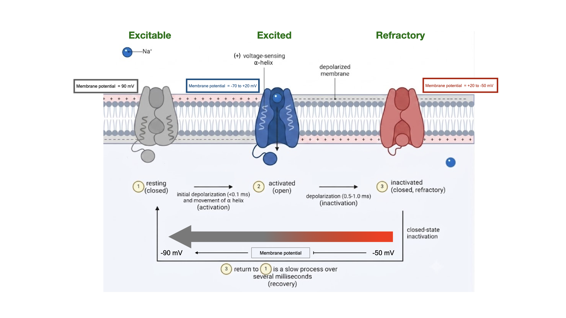
The fast Na⁺ channel changes conformation every beat. A drug’s effect therefore depends on when it binds and whether it leaves before the next impulse.
Resting · open · inactivated
Association + dissociation
How often the channel cycles
Phase-0 dV/dt · conduction · refractoriness
“Sodium-channel blocker” is not yet an explanation. State preference and recovery from block are what make Ia, Ib, and Ic behave differently.
Class I · Electrophysiologic effect · Post-repolarization refractoriness
The membrane-voltage clock and the channel-availability clock can separate.
ERP > APD90The membrane has completed 90% of its return toward resting potential. This defines a reproducible action-potential duration.
With Class I block, a sufficient fraction of Nav1.5 channels can remain drug-associated and unavailable. A premature impulse may therefore fail to propagate despite substantial repolarization.
ERP is defined operationally with a premature-stimulus test. It ends only when enough available fast Na⁺ current has recovered to support a propagated response.
Class Ia
Class Ib
Class Ic
Class II
Class III
Class IV
Foundations · 0.1
Visual context
Landmark trial · CAST-I
Cardiac Arrhythmia Suppression Trial · randomized, double-blind, placebo-controlled
Would suppressing ventricular ectopy (PVC) burden after myocardial infarction reduce sudden death?
Ventricular ectopy was suppressible during drug titration.
Encainide or flecainide versus matching placebo.
The active-drug arms were stopped early for excess mortality.
Relative risks in the preliminary CAST-I analysis
Final report: 63 deaths occurred with active treatment versus 26 with placebo; 43 versus 16 were arrhythmic deaths (P = .0004).